|
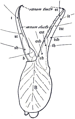
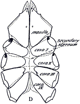
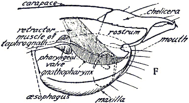
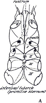
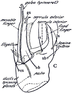
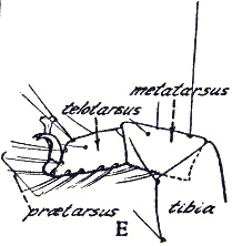
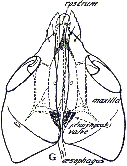
Fig. 4. Details of morphology, illustrating the terminology herein employed. A, coxal area of Chthonius ischnocheles (Hermann); B, exterior, lateral aspect of left chela of Geogarypus angulatus Chamberlin; C, exterior, lateral aspect of left chelicera of Garypus californicus Banks; D, coxal area and secondary sternum of Sternophorus sini Chamberlin; E, tarsus of Horus granulatus (Ellingsen); F, rostrum of Neobisium carolinensis (Banks), in place against the inner face of the left maxilla; G, ventral aspect of maxillae and rostrum of Hesperolpium slevini (Chamberlin).
|